The most amazing organ in our body is our “heart.” Since time immemorial, heart has been associated with many songs, poems, love and what not. If a person is in love, a sign of heart with an arrow mark shows the love. If a person is depressed in love, he/she is said to be heartbroken.
This amazing organ is always used to describe human emotions. The human heart is the essential organ of our body. So what is this heart? What does it do in our body? How does this heart function? Let us know more about the amazing facts about heart.
Where Is The Heart Located?
Heart is located in the center of the chest. It is present behind the breastbone or sternum. A part of the heart is slightly tilted towards the left side of the chest. It is located in the middle of the mediastinum at the level of thoracic T5-T8 vertebrae. A layer of membranous sac called pericardium surrounds the heart and this pericardium attaches to the mediastinum.
The front surface of the heart is behind the sternum and rib cartilages. The back surface of the heart is situated behind the vertebral column. The upper part of the heart is near the third costal cartilage and the lower part of the heart, which is the tip or apex of heart, is located to the left of the sternum between the fourth and fifth ribs.
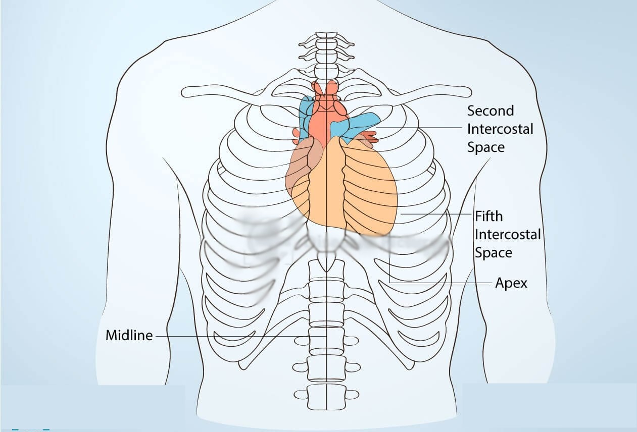
The heart is cone shaped and is the size of a fist. The base of the heart is facing upwards and the apex of this cone is downwards. Its largest part is offset to the left side of the chest. The left side of heart is stronger and larger since it pumps blood to all body parts.
The heart is located in between the lungs. The left lung is smaller than the right and has a cardiac notch in its border for the heart to fit there. An adult heart weighs about 250 to 350 grams.
What Are The Different Parts Of The Heart?
The human heart can be divided into:
- The four chambers of heart
- Muscular walls
- Blood vessels
- The conductive system
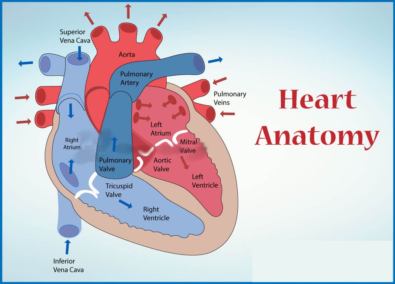
The Four Chambers Of Heart
Two upper chambers are called the atria (singular atrium)
- Right atrium
- Left atrium
Two lower chambers are called the ventricles
- Right ventricle
- Left ventricle
The Muscular Walls Of The Heart
The heart wall is made of three layers:
- Epicardium – The epicardium is the outer protective layer of the heart
- Myocardium- The myocardium is the middle muscular layer of the heart
- Endocardium – The endocardium is the inner layer of the heart
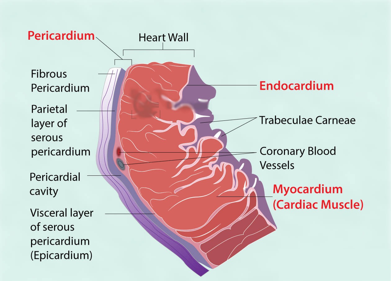
Epicardium
Epi means outer and cardium means heart, hence epicardium means the outer layer of the heart. It is also the inner visceral layer of the pericardium which attaches the heart to the mediastinum. The epicardium is made up of loose connective tissue and adipose (fat) tissue.
The function of epicardium is to protect the inner layers of heart and production of a fluid called pericardial fluid which helps reduce friction between the pericardial membranes. Epicardium also consists of coronary blood vessels which supply blood to the heart walls.
Myocardium
Myo means muscle, cardium means heart, myocardium means muscle layer of heart as this layer of heart contains the cardiac muscle fibers, which helps in contraction of heart. Myocardium is the middle layer of the heart. Myocardium is the thickest layer of the heart wall and the myocardium of the left ventricle is thicker than the rest as it requires power to pump oxygenated blood to all parts of our body.
Myocardium contains specialized type of muscle fibers which help in cardiac conduction. This bundle of fibers is made up of atrioventricular bundle and Purkinje fibers which carry electrical impulses that trigger the muscle fibers to contract.
Endocardium
“Endo” means inside or inner. Endocardium is the innermost layer of heart. It is a thin layer that covers the chambers and valves of the heart and is continuous with the endothelium of the large blood vessels. Infection of endocardium is called as endocarditis which can be a serious condition.
Blood Vessels Of Heart
The five major blood vessels of heart are
- Aorta
- Superior vena cava
- Inferior vena cava
- Pulmonary artery
- Pulmonary vein
The three main types of blood vessels are
- Arteries
- Capillaries
- Veins
The Aorta
The aorta is the largest artery in the body. It begins in the upper part of the left ventricle. The heart pumps blood into the aorta from the left ventricle through the aortic valve. The aorta is a tube like structure, an inch in diameter and about a foot long. It is again divided into four parts:
- Ascending aorta
- Aortic arch
- Descending thoracic aorta
- Abdominal aorta
The ascending aorta arises from the heart about 2 inches long. The coronary arteries are branches of ascending aorta supply the heart with blood. The aortic arch is a curved shape vessel over the heart which gives many branches that bring blood to head, neck and arms. The descending aorta goes down through the chest. Its branches supply blood to ribs and chest structures. The abdominal aorta starts at the diaphragm which branches to become iliac arteries of the abdomen and supplies blood to the major organs of the body.